



How the human eye works: anatomy and its individual parts

22 November 2026 · 9 min read
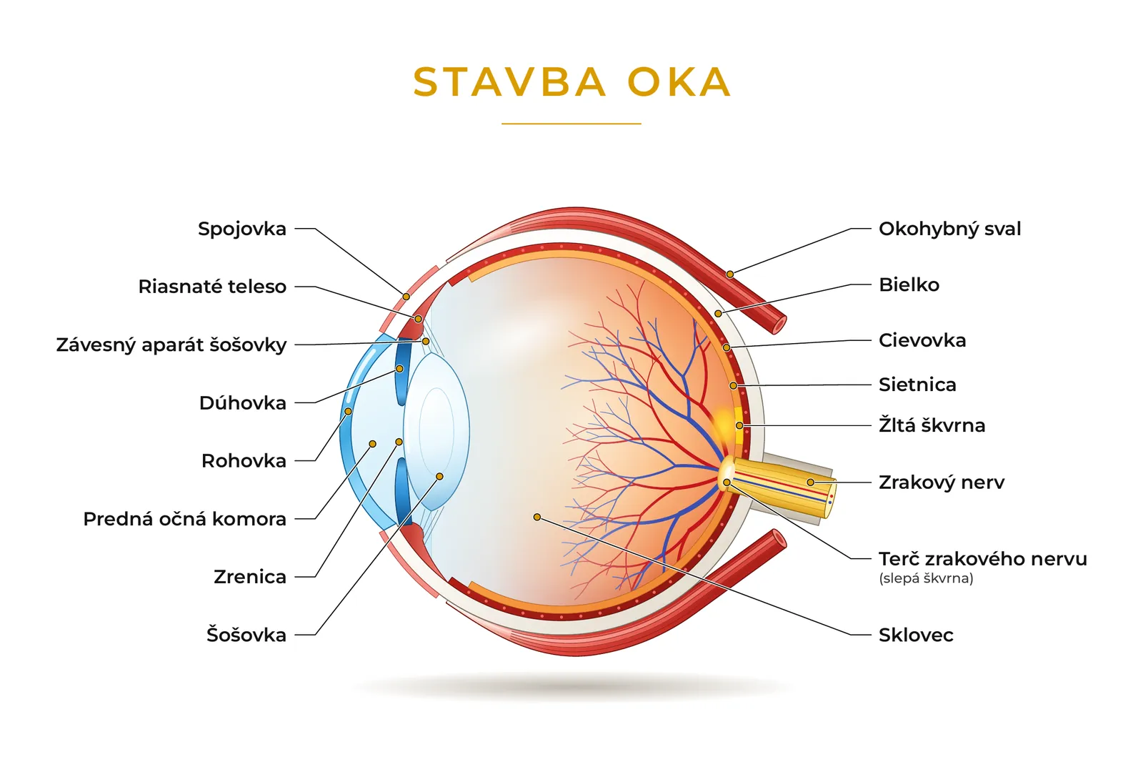
The eye is only about 24 millimetres across, yet it contains several layers, a lens, muscles and a nerve. They all have to work together precisely for you to see sharply. Once you understand what the eye is made of, it is easier to see why your vision is blurred, what the optometrist measures during an eye test and how they choose your glasses based on the measured prescription.
How the eye sees – in a nutshell
Light passes through the cornea, which refracts it the most. It then passes through the pupil – the opening in the iris – and the lens, which sharpens the image depending on whether you are looking into the distance or up close. Through the clear vitreous body it reaches the retina, where light is turned into a nerve signal. The optic nerve carries it to the brain, and only the brain assembles the image you “see”.
If light is not focused exactly on the retina, you see blurred. A spectacle lens with the right prescription helps focus the light in the right place. Depending on the type of error in the eye, we use a different type of lens:
- Myopia (poor distance vision) – corrected by a diverging lens, a lens with a minus prescription.
- Hyperopia (the eye strains, especially up close) – corrected by a converging lens, a lens with a plus prescription.
- Astigmatism (the image is blurred and distorted) – corrected by a cylindrical lens, which refracts light more strongly in one direction than in the other.
- Presbyopia (worse near vision after 40) – solved with reading glasses, progressive lenses (multifocal lenses) or office lenses.
In the animation, switch between how light passes through a healthy, a myopic and a hyperopic eye and what a spectacle lens does to it.
Front part of the eye
The front part of the eye receives light and directs it inside. It includes the cornea, conjunctiva, anterior chamber, iris and pupil.
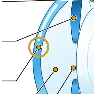
Cornea (lat. cornea)
A clear, curved layer right at the front – a kind of window of the eye. It refracts light more than any other part of the eye – about two thirds.
If the cornea is not evenly curved, astigmatism develops. The image is then blurred both at distance and near, and lines in one direction look sharper than in another. Contact lenses also sit on the cornea.
Conjunctiva (lat. tunica conjunctiva)
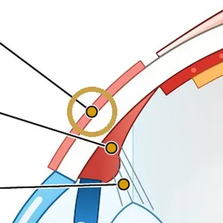
A thin, transparent membrane that covers the white of the eye and the inner side of the eyelids. It protects the eye and helps keep it moist.
When it becomes irritated or inflamed, the eye turns red, itches or waters – we call this conjunctivitis.
Anterior chamber (lat. camera anterior bulbi)
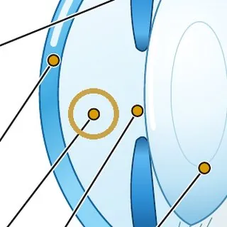
A small space between the cornea and the iris. It is filled with a clear fluid – the aqueous humour, which nourishes the front part of the eye.
The fluid is constantly produced and drained at the same time. Their balance determines the intraocular pressure. When the fluid does not drain sufficiently, the pressure rises and can damage the optic nerve.
Iris (lat. iris)
The coloured part of the eye. It is actually a muscle that changes the size of the opening in the middle – the pupil.
Why do we have different eye colours? The colour is determined by the amount of the pigment melanin in the iris. Brown eyes have a lot of it. Blue eyes have little, and there is actually no blue pigment in them at all – the blue colour is created by light scattering in the iris, much like the blue colour of the sky. Green and grey eyes are somewhere in between.
People with albinism have very little or no melanin in their body, including in the iris. The iris is therefore almost translucent and looks light blue to grey. In certain light or in a flash photo, the eyes can even look reddish or violet – light reflected from the retina, which is full of blood vessels, shines through the translucent iris. Because the iris cannot shade the eye well, their eyes are very sensitive to light. They often also have reduced visual acuity, because without melanin the retina does not fully develop at the point of sharp vision.
Pupil (lat. pupilla)
The black opening in the centre of the iris through which light enters the eye. It is black because the light that goes in hardly ever comes back out.
Why does the pupil narrow and widen? It works like the aperture of a camera and is controlled by a reflex – it happens by itself, without our will. In bright light it narrows to protect the retina from glare and make the image sharper. In dim light it widens to let as much light into the eye as possible. The pupil also narrows when looking up close.
The lens and focusing
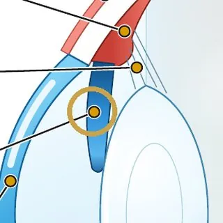
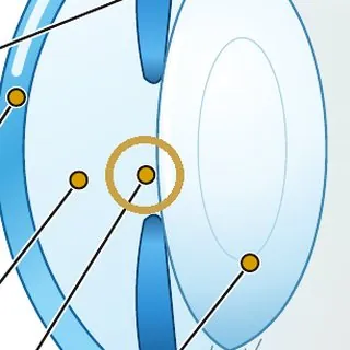
Thanks to the lens you can see sharply both at distance and up close. When focusing, it works together with a tiny muscle and fine fibres.
Lens (lat. lens)
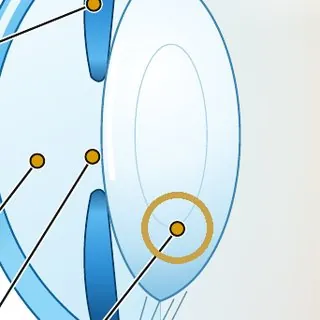
A clear, elastic body behind the pupil. It can change shape – become flatter or more curved – and so refocus the eye from distance to near. This ability is called accommodation.
When the lens becomes cloudy with age, a cataract develops.
Ciliary body (lat. corpus ciliare)
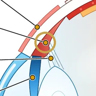
A ring of muscle around the lens. When the muscle contracts, the lens bulges and the eye focuses up close. When it relaxes, the lens flattens and the eye sees into the distance.
The ciliary body also produces the fluid that nourishes the front part of the eye.
Lens suspension (zonules) (lat. zonula ciliaris)
Hundreds of fine fibres from which the lens hangs as if on springs. They transfer the pull of the ciliary muscle to it – when they slacken, the lens bulges.
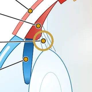
See how it all works in the animation:
With age the lens hardens and loses its elasticity. Around the age of 40 to 45, most people notice that they have to hold their phone or newspaper further away to read. This is presbyopia – a natural ageing of the eye, not a disease. The solution is reading glasses, office lenses for computer work or progressive (multifocal) lenses, which combine distance and near vision in one lens.
Back part of the eye
In the back part of the eye, light becomes an image. Light passes through the vitreous body, falls on the retina and the optic nerve carries it as a signal to the brain.
Vitreous body (lat. corpus vitreum)
A clear, jelly-like substance that fills most of the eye and keeps its round shape. Light passes through it unobstructed to the retina.
With age the vitreous gradually becomes more liquid. You may then notice “floaters” or cobwebs moving in your field of vision. They are mostly harmless. However, if many new floaters or flashes suddenly appear, see an eye doctor the same day.
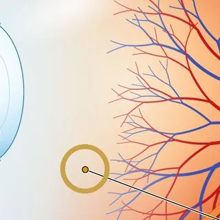
Retina (lat. retina)
A thin layer of nerve cells lining the inside of the eye. It works like the sensor in a camera – it turns light into a nerve signal.
It has two types of cells: rods, which let us see in dim light, and cones, which provide sharp and colour vision.
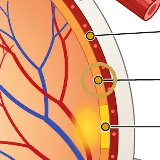
Rods and cones – two types of “sensors” in the retina (lat. bacilli et coni)
The retina contains two types of light-sensitive cells. Each of them does a different job.
Rods – vision in dim light
- There are about 120 million of them.
- They are very sensitive and need only a little light – thanks to them you can see at night.
- They do not distinguish colours, only shades of grey.
- They are mainly at the edges of the retina and detect movement and peripheral vision.
Cones – colour and sharpness
- There are about 6 million of them.
- They need more light and work mainly during the day.
- There are three types – for red, green and blue. By combining them you see all colours.
- Most of them are in the macula, which is why you see most sharply with it.
- “At night all cats are grey” – it's true. In low light only the rods work, and they cannot see colours.
- You will see a faint star in the night sky better if you look not directly at it but slightly to the side. Its light then falls on the rods at the edge of the retina.
- When you go from light into darkness, your eyes take up to 20–30 minutes to adjust. The rods have to “recharge”.
- Colour blindness occurs when one type of cone is missing or does not work properly. Most often red and green are confused, and it affects far more men than women.
Macula (yellow spot) (lat. macula lutea)
A small area in the centre of the retina with the highest number of cones. It is the point of sharpest vision – thanks to it you read, recognise faces and see details.
When the macula is damaged, for example by macular degeneration, a person stops seeing sharply right in the centre of the image.
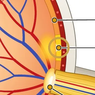
Optic disc (blind spot) (lat. discus nervi optici)
The place where the optic nerve leaves the retina. There are no light-sensitive cells here, so the eye sees nothing at this point. Normally you don't notice it – the brain fills in the gap and the other eye covers it.
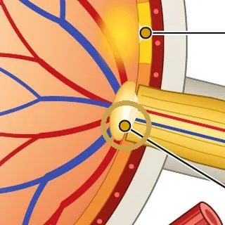
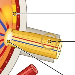
Optic nerve (lat. nervus opticus)
A kind of “cable” of more than a million nerve fibres that carries the signal from the retina to the brain. Only the brain assembles the image we see from it.
Damage to the optic nerve is irreversible. That is why there is so much talk about preventing glaucoma, which gradually destroys the nerve.
Outer layers and eye movement
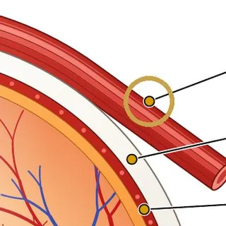
The eye is protected and nourished by three layers and moved by six muscles.
Sclera (white of the eye) (lat. sclera)
A firm white coat that protects the eye and keeps its shape. It is the white part of the eye you see in the mirror. At the front it merges smoothly into the clear cornea.
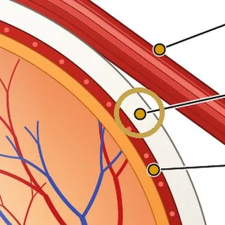
Choroid (lat. choroidea)
A thin layer rich in blood vessels between the sclera and the retina. It brings oxygen and nutrients into the eye and nourishes the retina.
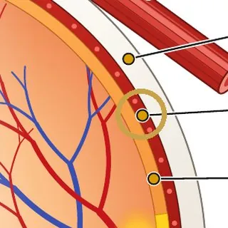
Extraocular muscles (lat. musculi bulbi)
Each eye has six muscles that move it up, down and sideways and also rotate it. They must work in perfect coordination so that both eyes look at the same point.
If they don't work together well, the eyes tire quickly, you get headaches or see double.
What this means for your glasses
The most common errors we correct with glasses are related precisely to where light is focused in the eye:
Myopia (short-sightedness) (lat. myopia)
The eye is slightly longer or refracts light too strongly, so the image is focused in front of the retina. You see poorly into the distance. A diverging lens helps – a lens with a minus prescription.
Hyperopia (long-sightedness) (lat. hypermetropia)
The image would only be focused behind the retina. The eye therefore has to strain constantly, especially when looking up close. A converging lens helps – a lens with a plus prescription.
Astigmatism (lat. astigmatismus)
The cornea is irregularly curved, so light is focused in different places in different directions. The image is blurred and distorted. A cylindrical lens helps.
Presbyopia (age-related long-sightedness) (lat. presbyopia)
With age the lens loses its elasticity and can no longer focus up close. After 40 you see your phone or reading material less well. Reading glasses, progressive lenses (multifocal lenses) or office lenses for computer work help.
During an eye examination we find out exactly how your eye focuses and suggest glasses that move the focus back onto the retina. We offer eye examinations at all 13 IMOOPTIK opticians.
At our opticians in Bratislava – Obchodná 57, Senica and Vrbové we have the DNEye® Scanner 2+ from the German company Rodenstock. The examination is contact-free and without the need for dilating eye drops. Remarkably, this device can reveal a lot of interesting things about the health of your eyes. Of course it does not replace an ophthalmologist, but during the examination we can detect early signs of disease and refer you to an eye doctor.
The device measures the eye at more than 7,000 points and, in addition to a precise prescription, it also determines:
- the shape and thickness of the cornea – reveals astigmatism and early signs of keratoconus,
- intraocular pressure – important for early detection of glaucoma,
- the depth of the anterior chamber – a narrow chamber carries a risk of an acute glaucoma attack,
- opacities in the eye – for example a beginning cataract,
- pupil size in daylight and at dusk – explains why some people see worse at night or when driving.
Frequently asked questions
Which part of the eye refracts light the most?
The cornea – it provides about two thirds of the eye's refractive power. The lens then fine-tunes the focus according to distance.
Why do I have a blind spot but can't see it?
There are no light-sensitive cells on the optic disc. The brain fills in the gap from the surroundings and the other eye covers it.
Are floaters dangerous?
Mostly not – they are natural changes in the vitreous body. However, if many new floaters, flashes or a curtain-like shadow suddenly appear, see an eye doctor the same day.
When did you last have your eyes checked?
Vision often deteriorates so slowly that you don't even notice it. We recommend an eye test every 1–2 years, and every year after 40.
- 13 opticians in western Slovakia
- Choose your appointment online
Founder of the IMOOPTIK network of opticians. He has worked in optics since 2009 – after finishing his studies he started at the GrandOptical opticians. In 2012 he opened his own optician's on Obchodná Street in Bratislava, and today IMOOPTIK runs 13 opticians in western Slovakia.