



6 common eye diseases: symptoms, how a diseased eye sees and when to see a doctor
22 November 2026 · 8 min read
Many eye diseases start unnoticed – they don’t hurt, and for a long time you may not realise your vision is getting worse. That is why it helps to know their signs and to have your eyes examined regularly. The illustrations below show what happens inside the eye with each disease and how vision can change.
1. Cataract (lat. cataracta)
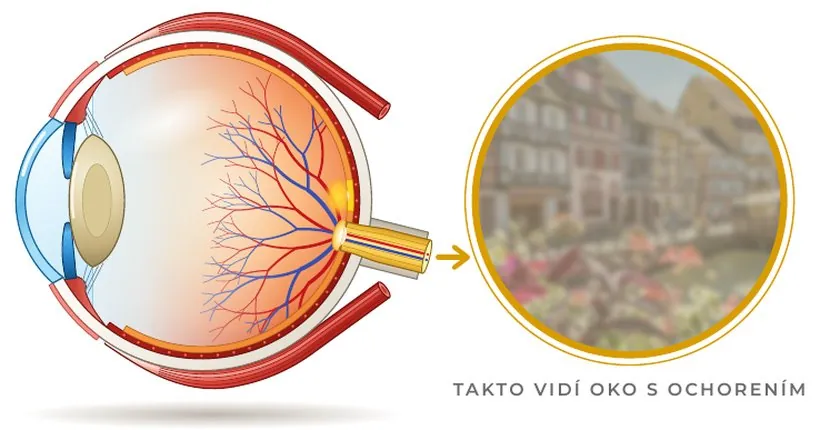
What happens: the lens inside the eye gradually becomes cloudy, most often due to age. Light passes through it less well and scatters.
What it looks like: vision is hazy, as if through a misted window, colours look faded, lights at night are dazzling and have halos around them. The prescription often changes quickly, so new glasses only help for a short time.
Treatment: cataract is treated by surgery – the eye doctor replaces the cloudy lens with an artificial one. It is one of the most common and safest eye operations. After surgery you usually need new glasses, especially for near vision.
2. Glaucoma (lat. glaucoma)
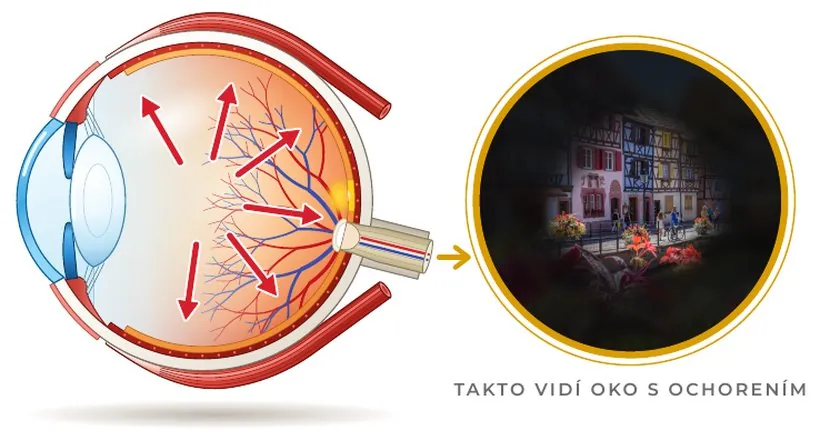
What happens: the optic nerve is gradually damaged, often due to increased intraocular pressure. Slovak ophthalmologists describe it as the second most common cause of blindness.
What it looks like: the field of vision narrows from the edges – as if you were looking through a tube. The insidious thing is that glaucoma doesn’t hurt and you may not notice the first symptoms at all.
Who is at risk: people over 40, with glaucoma in the family, higher intraocular pressure, high myopia, diabetes or vascular disease.
Treatment: nerve damage cannot be reversed, but its progression can be stopped or slowed – with eye drops, laser or surgery. Prevention is therefore key: after 40, doctors recommend a preventive check-up with an eye doctor once a year.
3. Age-related macular degeneration (AMD) (lat. degeneratio maculae)
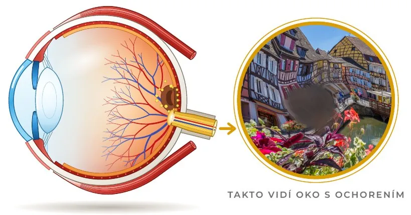
What happens: the macula – the point of sharpest vision in the centre of the retina – becomes damaged. It mainly affects people over 50.
What it looks like: a dark or blurred spot appears in the centre of the image, straight lines look wavy, reading and recognising faces becomes difficult. The edges of the field of vision remain intact.
Treatment: the wet form is treated by an eye doctor with injections into the eye, the dry form is monitored regularly. Smoking greatly increases the risk. Magnifying aids also help people with low vision.
- If you wear reading glasses, put them on.
- Hold your phone, or sit, about 30–40 cm from the screen.
- Cover one eye and look at the red dot in the centre.
- Then repeat with the other eye.
The test is only indicative and does not replace an examination.
4. Diabetic retinopathy (lat. retinopathia diabetica)
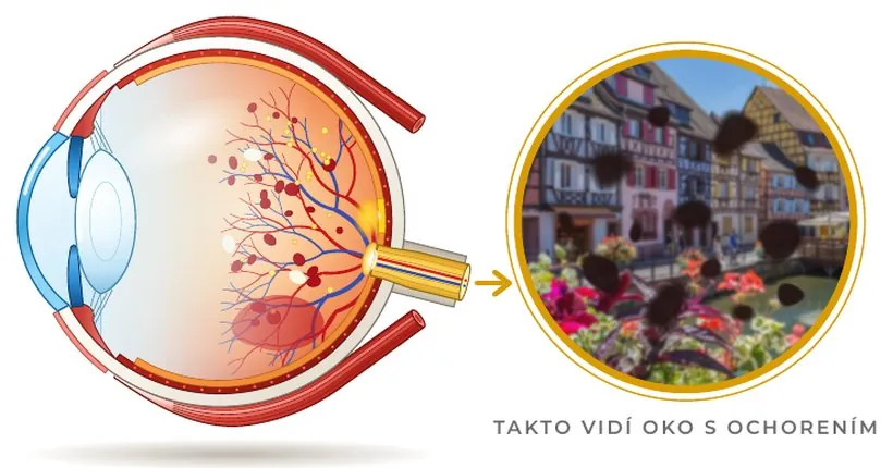
What happens: long-term high blood sugar damages the tiny blood vessels in the retina. The vessels can leak or bleed, and new, fragile vessels may form.
What it looks like: often no symptoms at first, later dark spots and blurred, fluctuating vision.
Treatment: every person with diabetes should see an eye doctor regularly for a fundus examination, even if they see well. Well-controlled blood sugar and blood pressure greatly reduce the risk. If your prescription changes often because of diabetes, mention it during your eye test at the optician’s too.
5. Retinal detachment (lat. ablatio retinae)
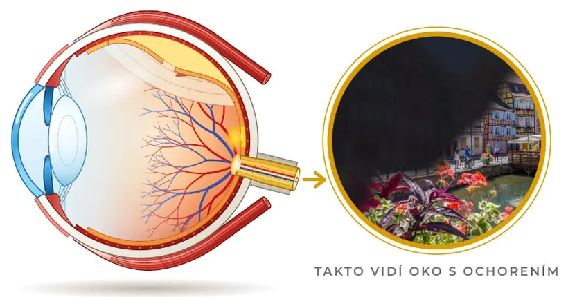
What happens: the retina separates from the choroid, which nourishes it. This is an emergency that requires immediate treatment.
What it looks like: suddenly many new floaters, flashes of light in one or both eyes and a dark shadow or “curtain” in the field of vision. It doesn’t hurt.
Who is at risk: people with high myopia, after an eye injury, after cataract surgery or with retinal detachment in the family.
Treatment: with these symptoms you need to see an eye doctor or go to the emergency department straight away – early treatment can save your sight. It is treated with laser or surgery.
6. Keratoconus (lat. keratoconus)
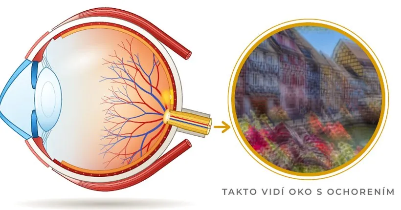
What happens: the cornea thins and bulges into a cone shape. It most often begins at a young age.
What it looks like: blurred, distorted and double vision, increasing astigmatism that becomes harder and harder to correct with glasses.
Treatment: in the early stage glasses are enough, later special rigid contact lenses. An eye doctor can slow down or stop the disease with a procedure called cross-linking. In advanced cases a corneal transplant is performed. If your astigmatism is getting worse quickly, have it checked.
What role does the optician play
An optometrist at the optician’s does not treat eye diseases – that is the job of an eye doctor. During an eye examination, however, they are often the first to notice that something is not right. A warning sign is, for example, an unexpectedly rapid change in prescription, or vision that cannot be sharpened with glasses. In such a case we will refer you to an eye doctor.
DNEye® Scanner
A device from the German company Rodenstock measures the eye at more than 7,000 points. Contact-free and without the need for dilating eye drops, it measures:
- precise prescription to hundredths of a dioptre and subtle optical aberrations of the eye for distance and near,
- a map and the thickness of the cornea – helps detect keratoconus,
- the depth of the anterior chamber – a narrow chamber increases the risk of glaucoma,
- intraocular pressure,
- pupil size in daylight and at dusk,
- opacities in the eye – for example a beginning cataract.
From the measured data we can also make Rodenstock biometric lenses, calculated precisely for your eye.
More about DNEye® →Visionix VX120+
A multi-diagnostic device that, contact-free and without the need for dilating eye drops, measures:
- precise prescription and subtle optical aberrations of the eye,
- a map and the thickness of the cornea – helps detect keratoconus,
- the depth of the anterior chamber and the chamber angle – a narrow angle increases the risk of glaucoma,
- intraocular pressure,
- pupil size under different lighting,
- opacities in the eye – for example a beginning cataract.
However, it does not support the production of biometric lenses.
A diagnosis is always made by an eye doctor. We offer eye examinations at all 13 IMOOPTIK opticians.
Frequently asked questions
What is the difference between cataract and glaucoma?
A cataract is a clouding of the lens – it is treated by surgery and vision is usually restored. Glaucoma is damage to the optic nerve – lost vision cannot be restored, treatment only stops the disease.
How often should I have my eyes checked?
Adults without problems should have an eye test every 1–2 years. After 40, with diabetes or eye disease in the family, also a preventive check-up with an eye doctor once a year.
Can you detect an eye disease during an examination at the optician’s?
A diagnosis is always made by an eye doctor. During an eye test, however, we can notice warning signs and recommend that you see a doctor. At six of our opticians we also have DNEye® Scanner devices (Bratislava – Obchodná 57, Senica, Vrbové) and Visionix VX120+ (Komárno, Bratislava – Miletičova, Karlova Ves), which, contact-free and without dilating drops, also measure intraocular pressure, the cornea and opacities in the eye.
The illustrations and vision simulations are schematic and do not replace an examination. This article is for information only. If your vision changes, see an eye doctor or optometrist.
When did you last have your eyes checked?
Vision often deteriorates so slowly that you don’t even notice it. We recommend an eye test every 1–2 years, and every year after 40.
- 13 opticians in western Slovakia
- Choose your appointment online
Founder of the IMOOPTIK network of opticians. He has worked in optics since 2009 – after finishing his studies he started at the GrandOptical opticians. In 2012 he opened his own optician’s on Obchodná Street in Bratislava, and today IMOOPTIK runs 13 opticians in western Slovakia.